Ultrasound
The use of Ultrasonic products is increasing as new techniques and improvements in instrument performance constantly expand the range of applications. Spectrum digitizers are ideal tools for making ultrasonic measurements and can play a key role required in the development, testing and operation of these products. Spectrum digitizers and arbitrary waveform generators offer a wide range of bandwidths, sampling rates, and dynamic range to match the broad spectrum of ultrasonic measurement needs. When wide dynamic range and maximum sensitivity is required high-resolution 14 and 16 bit digitizers are available for capturing and analyzing ultrasonic signals from 100 kHz up to 250 MHz in frequency. A cost effective range of 8 bit digitizers is also available to cover frequency ranges from 5 MHz up to 1.5 GHz. Typical ultrasound applications include Non-Destructive Testing (NDT), Ultrasonic Testing (UT), Doppler Effect Flow-meters, Time-of-flight Diffraction (TOFD), Range Finding, Scanning Acoustic Microscopy (SAM) and Tomography (SAT), Medical Sonography and Ultrasonography, Phased array ultrasonics, Laser ultrasonics and Acoustic Emission.
Spectrum Product Features
- 12, 14 and 16 Bit Resolution
- Segmented Memory with FIFO Readout
- Low Dead-Time between triggers (< 80 ns)
Matching Card Families
44xx
Family
A/D family
Sample rate
130 MS/s - 400 MS/s
Resolution
14 Bit 16 Bit
22xx
Family
A/D family
Sample rate
1.25 GS/s - 5 GS/s
Resolution
8 Bit
59xx
Family
A/D family
Sample rate
5 MS/s - 125 MS/s
Resolution
16 Bit
33xx
Family
A/D family
Sample rate
6.40 GS/s - 10 GS/s
Resolution
12 Bit
Related Documents
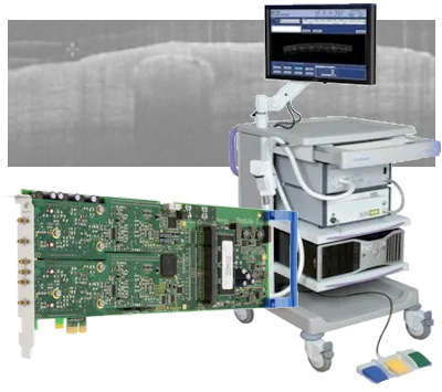
Case Study: OCT application for skin cancer diagnosis
The VivoSight® OCT scanner uses the technique of swept-source Optical Coherence Tomography (SS-OCT) for cross-sectional imaging of skin. This is a significant new tool to assist in diagnosis and treatment of skin cancers and other skin conditions.
Ultrasonic Applications
The use of Ultrasonic products is increasing as new techniques and improvements in instrument performance constantly expand the range of applications. Spectrum digitizers are ideal tools for making ultrasonic measurements and can play a key role required in the development, testing and operation of these products. Spectrum digitizers and arbitrary waveform generators offer a wide range of bandwidths, sampling rates, and dynamic range to match the broad spectrum of ultrasonic measurement needs
Research Papers
Micro Bubbles Cavitation
The Shanghai Jiao Tong University in China is conducting research into the properties of daughter bubbles that are generated by inertial cavitation of preformed microbubbles. For detection of inertial cavitation and scattering, transducers produce signals that are acquired by an M4i.4410-x8 130 MS/s, 16-bit Digitizer. The data is then transferred to a computer for Fourier transform and power spectrum analysis. A white paper discussing this ultrasonics sonochemistry research is available for download
Research PaperPhotoacoustic Wavefront Shaping
At London’s St Thomas’ Hospital in the UK, they are working on a high-speed photoacoustic-guided wavefront shaping method, that uses a relatively simple experimental setup, with potential for in vivo applications. Part of the system uses an M4i.4420-x8 250 MS/s, 16-bit Digitizer to acquire ultrasonic signals. The full research paper discussing the experimental setup and results can be found here:
Research PaperUltrasound Endoscope
The College of Biophotonics, South China Normal University, China, is improving the performance of photoacoustic/ultrasound endoscopes. Their research uses an M4i.4420-x8 250 MS/s, 16-bit Digitizer as part of dual-modality endoscope. A research paper shows the potential for using the tens-of-micron-resolved PA/US endoscope for in vivo anatomical imaging in the clinical detection of colorectal diseases.
Research PaperDrug Delivery for Cancer Treatment
The University of Leeds and the Leeds Institute of Medical Research, Leeds, U.K. are studying the use of nanobubbles as a drug delivery agent for cancer treatment. A Spectrum M4i.4420-x8, 250 MS/s, 16-bit Digitizer is used as the data acquisition card that collect acoustic emission signals. A paper discussing the research can be found here:
Research PaperHandheld Photoacoustic Microscopy
The MOE Key Laboratory of Laser Life Science and Institute of Laser Life Science, at the South China Normal University, in China has developed a Photoacoustic Imaging (PAI) pen that can be handheld (performing forward detection and lateral detection) to extend the application of photoacoustic (PA) microscopy to areas such as the oral cavity, throat, cervix, and abdominal viscera. The experimental setup uses an M4i.4450-x8 500 MS/s, 14 bit, digitizer to acquire the sensor signals. A paper discussing the PAI pen and the test results can be found here:
Research PaperLarge Area all-optical Ultrasound Imaging
The University College London has developed a method for large area all-optical ultrasound imaging using robotic control that involves the use of an M4i.4420-x8 250 MS/s, 16-bit digitizer. A white paper on the development can be found here:
Research PaperPhotoacoustic Imaging
Find out how Nanyang Technological University, Singapore, uses a high speed 16 bit Spectrum digitizer M4i.4420-x8 for Photoacoustic Imaging by clicking here:
Research PaperUltrasonic 3D Endoscopic Imaging System
See how the Department of Medical Physics and Biomedical Engineering, at University College London, UK, use an M4i.4420-x8 high-resolution digitizer in a miniature all optical ultrasonic 3D endoscopic imaging system by clicking here:
Research PaperNon-Invasive Cancerous Tissue Treatment
Click below to find out how the School of Electronic and Electrical Engineering, University of Leeds, UK, are using a Spectrum M4i.4420-x8 high-resolution digitizer, plasmonic gold nanorods and high intensity focused ultrasound (HIFU) to improve non-invasive techniques for the treatment of cancerous tissue
Research PaperTherapeutic Ultrasound Brain Deseases Treamtment
At the Queensland Brain Institute, University of Queensland, researchers are using a Spectrum M4i.4421-x8 16 bit digitizer to study ultrasound propagation in materials that are used to modell the human skull as well as investigating therapeutic ultrasound as a potential means to treat deseases of the brain.
Research PaperBreast Ultrasound Scanning System
The Laboratory of Acoustical Waveform Imaging, Department of Imaging Physics, Delft University of Technology, Delft, The Netherlands, is developing the Delft Breast Ultrasound Scanning System (DBUS) as a means for detecting the presence of tumors. Find out how they are using the 14 bit M3i.4142 digitizer by following this link:
Research PaperReal-Time 2D Ultrasound Imaging
A video-rate all-optical ultrasound imaging system, where ultrasound is generated and detected using light, has been demonstrated at the Department of Medical Physics and Biomedical Engineering, University College London, UK. The system uses a Spectrum M4i.4420-x8 high resolution 16 bit digitizer to acquire signals from a broadband photodiode. Details on how the system enables real-time, video-rate 2D ultrasound imaging, at a frame rate of 15 Hz, can be found here:
Research PaperOptical Resolution Photoacoustic Microscopy
The School of Biomedical Engineering, Tohoku University, Japan is using a 5 GS/s M4i.2230-x8 Digitizer to achieve optical resolution photoacoustic microscopy with sub-micron lateral resolution for visualization of cells and their structures. Details and results of their experimental setup can be found here:
Research PaperAcoustic-Resolution-based Photoacoustic Microscope (ARPAM)
At the Central South University in Changsha, China, they are using an M4i.2233-x8 high speed digitizer in an improved acoustic-resolution-based photoacoustic microscope (ARPAM) that helps to resolve the conflict between lateral resolution and depth of field. The article is available here:
Research Paper3 Megapixel Ultrasonic Microscope
The University of Helsinki, Finland, has developed a 3 Megapixel Ultrasonic Microscope using a Spectrum M4i.6631-x8 AWG for signal transmission and an M4i.2233-x8 digitizer for data acquisition. IEEE members can download the full article here:
Research PaperUltrasonic Velocity in Ultra-Low Expansion Glass
The temperature dependence of ultrasonic velocity in ultra-low expansion (ULE) glass is being studied at the Institute of Optics and Electronics, Chinese Academy of Sciences, Chengdu, China. In a white paper they present a correlation method to determine the ultrasonic TOF in ULE glass and to further obtain the ultrasonic longitudinal wave velocity indirectly. The experimental setup uses an M4i.2220-x8 2.5 GS/s, 8-bit Digitizer to capture signals from an ultrasonic pulser/receiver.
White PaperStable Cavitation Activity of circulating Microbubbles
At the School of Sensing Science and Engineering, Shanghai Jiao Tong University, in China they are using a closed-loop feedback controller, based on pulse length (PL), regulation method to improve the temporal stability of stable cavitation (SC) activity. The aim is to achieve controllable and desirable SC activity in target regions for improved therapeutic efficiency and biosafety. A research paper showing the improvements obtained can be found here 7 The setup uses an M4i.4410-x8 130 MS/s, 16-bit PCIe Digitizer to determine the acoustic emission properties of the circulating microbubbles. The digitizer acquires signals produced by a transducer and passes the data at high speed to a computer for subsequent processing.
Reserach PaperDaughter- and Microbubble Studies
Inertial cavitation (IC) of the preformed microbubbles is being investigated for ultrasound imaging and therapeutic applications. However, microbubbles rupture during IC, creating smaller daughter bubbles (DBs), which may cause undesired bioeffects in the target region. The School of Sensing Science and Engineering, Shanghai Jiao Tong University, in China, is researching the properties of the daughter bubbles with the aim of achieving controllable cavitation activity. A research paper on the topic can be found here with the experimental setup using an M4i.4410-x8 130 MS/s, 16-bit Digitizer to acquire the relevant transducer signals
Research PaperHigh-Order Pulse-Echo Ultrasound
The Institute for Biomedical Engineering, Department of Information Technology and Electrical Engineering, ETH Zurich, Switzerland they are researching ways to improve scanning acoustic microscopy by introducing high order reflection pulse-echo (HOPE)-ultrasound. HOPE is a novel method that leverages high order reflections to improve on several aspects of conventional ultrasound imaging. A research paper on the HOPE development, which uses an M4i.4420-x8 250 MS/s, 16-bit Digitizer, to acquire the related transducer signals, can be found here
Research PapersAll-Optical Ultrasound Imaging
All-optical ultrasound (AOUS) imaging, uses light to both generate and detect ultrasound, and is an emerging alternative to conventional electronic ultrasound imaging. At the Department of Medical Physics & Biomedical Engineering, University College London, United Kingdom, they've developed the first AOUS system with a flexible, handheld imaging probe, which represents a critical step towards clinical translation. White papers describing the research, which uses the M4i.4420-x8, 250 MS/s, 16-bit Digitizer for signal acquisition, are available here:
White PaperAgeing Dementia Research
At the Queensland Brain Institute, The University of Queensland, in Australia they are studying possible ways to treat Alzheimer's disease using scanning ultrasound. A paper discussing the research, which uses an M4i.4421-x8 250 MS/s, 16-bit Digitizer for signal acquisition, can be found here:
Research PaperTesting Concrete Structures
onitoring of the behaviour of concrete under load is an important task in the safety evaluation of concrete structures. At the College of Automotive and Mechanical Engineering, Changsha University of Science and Technology, in China, they are applying the coda wave method to the non-destructive testing of concrete structures and looking how it can improve measurement penetration and sensitivity. A research paper on their work, which uses an M4i.4420-x8 250 MS/s, 16-bit Digitizer for signal acquisition, can be found here:
Research PaperAcoustic horn characterization
The National Defense Academy, in Japan are investigating source of acoustic cavitation noise from bubble clusters under ultrasonic horns. Their research uses a hydrophone and schlieren visualization together with an M4i.4451-x8 500 MS/s, 14-bit Digitizer to help determine the horns characteristics for direction, autocorrelation and frequency. A research paper discussing the work can be found here:
Research Paper3D Photoacoustic Imaging
The School of Engineering Medicine, Beihang University, Beijing, China have developed a technique for compressed single-shot 3D photoacoustic imaging with a single-element transducer. They setup uses an M2p.5961-x4 125 MS/s, 16-bit Digitizer to collect the transducer signals as outlined in the reference paper below
Reference Paper